When is arthroscopy useful?
In hip dysplasia, arthroscopy alone is usually not the right treatment: removing acetabular rim can make the joint less stable. We therefore assess the shape of the whole hip before every arthroscopy.
- Femoroacetabular impingement (FAI), cam or pincer type
- Tears of the labrum
- Localised cartilage damage and loose bodies in the joint
How do we reach the joint?
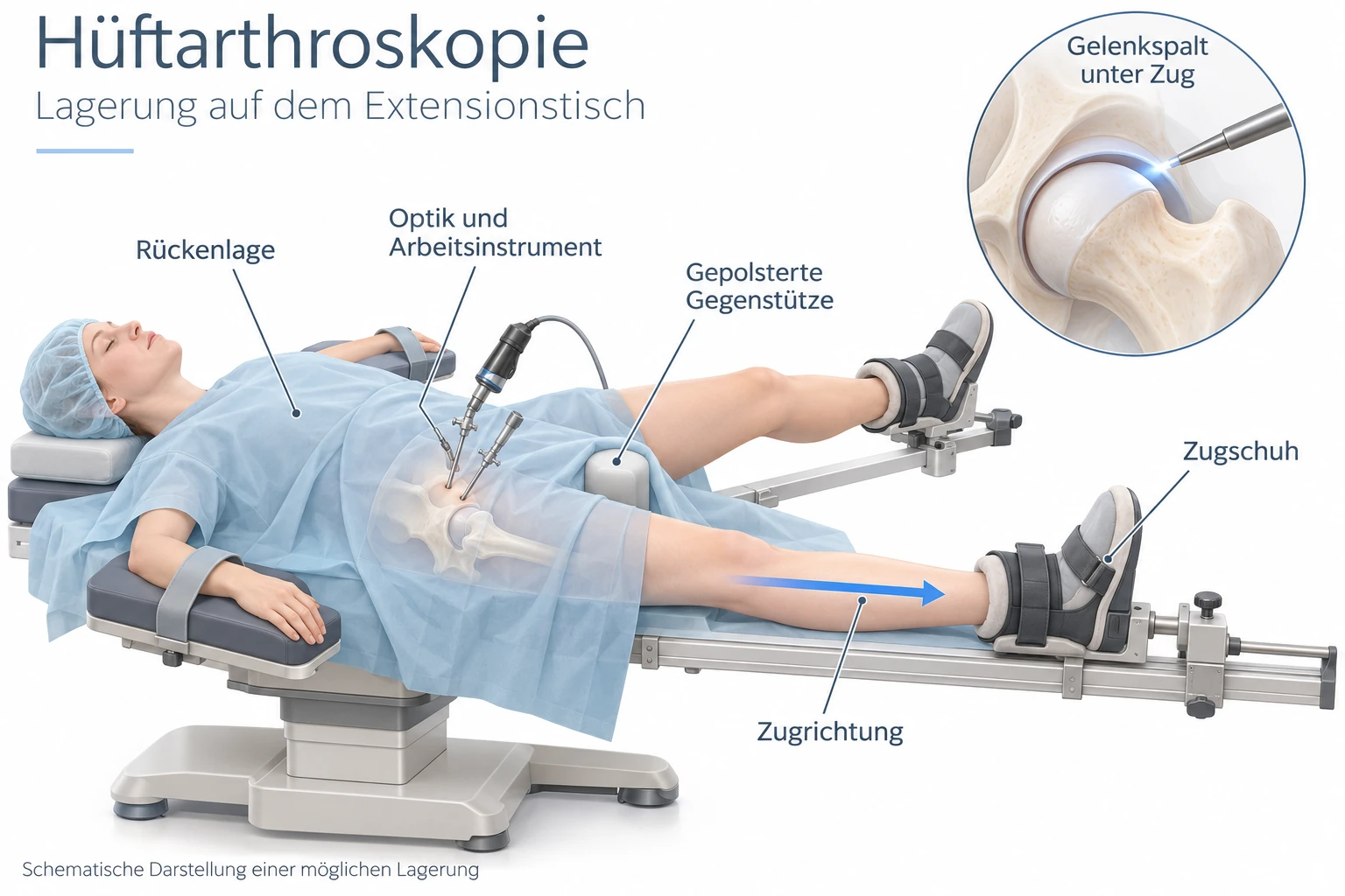
To reach the hip joint with the camera and fine instruments, the joint is carefully distracted during the operation (traction). The schematic illustration shows the positioning on the traction table and the space this creates between the femoral head and socket.
What is done?
The steps needed depend on the shape of your hip and the condition inside the joint. We often combine several steps in one operation.
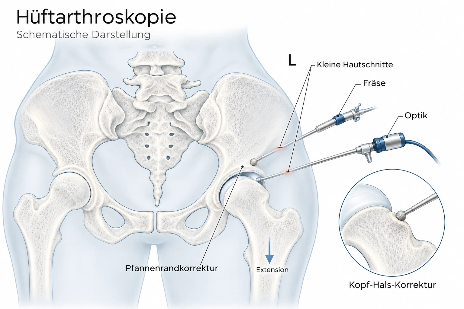
Cam correction (femoral head–neck reshaping)
In cam impingement, the transition from the femoral head to the femoral neck is not sufficiently slender. A bony bump moves beneath the acetabular rim during flexion. This shears the cartilage from within and tears the labrum.
We remove this bump with a fine burr until the transition is round again. Using X-ray guidance and movement checks during the operation, we make sure that enough bone is removed. At the same time, not too much must be removed so that the femoral neck remains stable. Whether the cam correction is complete is a major factor in the outcome.
Pincer correction (acetabular rim correction)
In pincer impingement, the socket covers the femoral head too much in one area. The femoral neck hits the acetabular rim early.
To correct this, we temporarily detach the labrum from the bone, trim the protruding rim by a few millimetres and then reattach the labrum. We deliberately correct conservatively: excessive trimming makes the joint unstable. We therefore measure the socket coverage precisely before the operation. If the entire socket is malpositioned, for example tilted backwards (retroversion), correction at the pelvis is sometimes the better option.
Labral repair (refixation of the labrum)
The labrum acts like a seal. It keeps synovial fluid in the joint, creates slight negative pressure and thereby stabilises the femoral head in the socket.
If it is torn or detached, we sew it back to the acetabular rim using small suture anchors. The anchors are a few millimetres in size and remain in the bone. Studies show that reattachment produces better results than simply removing the labrum. We therefore always try to preserve it.
Labral reconstruction
If the labrum is so badly damaged or deficient that repair is no longer possible, we replace it with tendon tissue from your own body (autograft). This restores the seal of the joint. Reconstruction is more complex and aftercare takes a little longer.
Cartilage and loose bodies
We remove loose pieces of cartilage or bone. Small, localised cartilage defects are smoothed or treated so that new replacement tissue can form in the defect. Extensive cartilage damage, by contrast, cannot be treated adequately by arthroscopy.
Closure of the joint capsule
At the end, we usually close the joint capsule with sutures. This preserves the natural stability of the hip.
After the procedure
Aftercare is just as important for the outcome as the operation itself. It takes place in phases. How quickly you progress depends on what was operated on.
In hospital
- You usually stay for one or two nights.
- You get up with physiotherapy and forearm crutches on the day of surgery or the first day afterwards.
- You receive a stepped pain management plan. Mild pain in the groin and buttock is normal during the first few days.
- Numbness on the outer thigh or in the perineal area can occur because of positioning and usually resolves within days to weeks.
Phase 1 – Protection (approximately weeks 1 to 3)
Weight bearing: Partial weight bearing with forearm crutches, usually at around 20 kg. After cam or pincer correction alone without labral repair, weight bearing can often be increased more quickly. After labral reconstruction or cartilage treatment, you maintain partial weight bearing for longer, usually up to six weeks.
Movement: Do not bend the hip beyond 90° during the first few weeks; do not sit low or squat. Avoid strong rotational movements and extending the leg backwards.
Exercise bike: Cycling on an exercise bike without resistance and with the saddle set high is usually allowed from the first few days. It keeps the joint mobile and helps prevent adhesions.
Thrombosis prevention: You receive thrombosis prevention for the period of partial weight bearing.
Calcification: To protect against calcification in the tissue (heterotopic ossification), we often prescribe an anti-inflammatory medicine for a short time. Studies show that this significantly reduces the risk.
Phase 2 – Mobility and increasing weight bearing (approximately weeks 3 to 6)
You gradually increase weight bearing up to full weight bearing and progressively stop using crutches. The prerequisite is a safe gait without limping.
The hip becomes increasingly mobile.
You begin targeted strengthening of the buttock and core muscles, initially without weights.
Swimming is possible once wound healing is complete. You should avoid breaststroke initially.
Phase 3 – Strength and stability (approximately weeks 6 to 12)
You build strength in a targeted way and train balance and leg alignment control.
Training in the gym with gradually increasing load is now possible.
For returning to work: office work is often possible again after two to three weeks, and heavy physical work usually after six to twelve weeks.
Phase 4 – Return to sport (from approximately month 3)
Jogging: usually after around three months, provided strength and mobility are sufficient.
Sports involving jumping, sprinting, changes of direction or body contact (for example football, tennis, handball and martial arts): usually after four to six months.
Function rather than the calendar: What matters is that you are pain-free and pass strength and stability tests. Your physiotherapist checks this with you.
Please be patient: it usually takes six to twelve months to reach the final result. Periods of mild symptoms can occur in between, for example when you increase your activity. This is normal. You receive your personal instructions, adapted to the steps performed, in your discharge letter.
Please contact us if you develop a fever, increasing redness or swelling of the wound, swelling of the calf, or new burning nerve pain.
What you can expect – what studies show
Arthroscopy or physiotherapy? Two large randomised studies from the UK (FASHIoN, Lancet 2018; FAIT, BMJ 2019) compared arthroscopy with targeted physiotherapy for hip impingement. After surgery, symptoms and quality of life were significantly better on average. Physiotherapy nevertheless remains a useful first step, especially for mild symptoms.
Preserving the labrum is worthwhile: Repairing the labrum rather than removing it produces better results. This is shown by both randomised studies and long-term observations.
Return to sport: In review articles, the large majority of athletes return to their sport, many at their previous level.
Cartilage is decisive: The best results are achieved by patients whose cartilage is still well preserved at the time of surgery. With advanced osteoarthritis, the benefit is small and the risk of eventually needing an artificial joint increases significantly.
The correct diagnosis is crucial: The most common cause of a poor outcome is an unrecognised or inadequately corrected shape abnormality, such as missed dysplasia, a remaining cam bump or an abnormal rotation of the femur. We therefore take considerable time for diagnosis before surgery.